Home
/ Svt Ekg / Supraventricular Tachycardia Svt Ecg Simulator Arrhythmia Simulator Youtube : This abnormal accessory pathway allows the
Svt Ekg / Supraventricular Tachycardia Svt Ecg Simulator Arrhythmia Simulator Youtube : This abnormal accessory pathway allows the
Svt Ekg / Supraventricular Tachycardia Svt Ecg Simulator Arrhythmia Simulator Youtube : This abnormal accessory pathway allows the. From wikipedia, the free encyclopedia supraventricular tachycardia (svt) is an abnormally fast heart rhythm arising from improper electrical activity in the upper part of the heart. The second ecg is a patient who was initially thought to be in svt. In all other svts, they precede the qrs, if ps are present. Ecg library homepage regular broad complex tachycardias can be ventricular (vt) or supraventricular (svt with aberrancy) in origin, and differentiation between the two will significantly influence your management of the patient. A normal heart rate is 60 to 100 beats per minute.
A heart rate of 150 should make you suspect atrial flutter is present. Often there is 2:1 conduction to the ventricle, giving a ventricular heart rate of approximately 150 bpm (as in this ecg). Ecg library homepage regular broad complex tachycardias can be ventricular (vt) or supraventricular (svt with aberrancy) in origin, and differentiation between the two will significantly influence your management of the patient. Supraventricular tachycardia is an abnormal rhythm with a very fast heart rate (140 to 240 bpm). To catch an episode, your doctor may give you an ecg monitor to wear at home that will record your heart rhythm over time.
How My Watch Helped Get A Diagnosis Applewatch from i.imgur.com The first ecg below was mistaken for svt and it's easy to see why. Unfortunately, the electrocardiographic differentiation of vt from svt with aberrancy is not always possible. Supraventricular tachycardia (svt) is as an abnormally fast or erratic heartbeat that affects the heart's upper chambers. The ekg/ecg typically is a narrow complex tachycardia with a rate above 120 bpm. Look for r and r'(prime) (only in v1 and v2 or v5 and v6) 3. Inverted p waves are sometimes seen after the qrs complex. Svt can also cause electrical alternans that disappears when sinus rhythm is restored Describes ecg findings for common types of svt.
Inverted p waves are sometimes seen after the qrs complex.
Another cause of psvt can be when there are two pathways between the av node and the ventricles. Use these ekg practice tests to help you become proficient in your rapid rhythm identification. In svts with rapid ventricular rates, p waves are often obscured by the t waves, but may be seen as a hump on the t. The term has latin roots. Ecg library homepage regular broad complex tachycardias can be ventricular (vt) or supraventricular (svt with aberrancy) in origin, and differentiation between the two will significantly influence your management of the patient. Supraventricular tachycardia (svt) is as an abnormally fast or erratic heartbeat that affects the heart's upper chambers. Svt is also called paroxysmal supraventricular tachycardia. Supraventricular tachycardia is an abnormal rhythm with a very fast heart rate (140 to 240 bpm). The ekg/ecg typically is a narrow complex tachycardia with a rate above 120 bpm. Ekg practice test 1 this ekg practice test is designed to help you learn to recoginze all of the ekg rhythms that you will encounter during emergencies and during the aha acls provider course. A normal heart rate is 60 to 100 beats per minute. Supraventricular tachycardia is a rapid heart rate (tachycardia, or a heart rate above 100 beats per minute) that is caused by electrical impulses that originate above the heart's ventricles. From wikipedia, the free encyclopedia supraventricular tachycardia (svt) is an abnormally fast heart rhythm arising from improper electrical activity in the upper part of the heart.
Supraventricular tachycardia (svt) svt is a broad term for a number of tachyarrhythmias that originate above the ventricular electrical conduction system (purkinje fibers). Sinus tachycardia has a rate of 100 to 150 beats per minute and svt has a rate of 151 to 250 beats per minute. Once the patient was given adenosine, flutter waves were revealed. The ekg/ecg typically is a narrow complex tachycardia with a rate above 120 bpm. Svt can also cause electrical alternans that disappears when sinus rhythm is restored
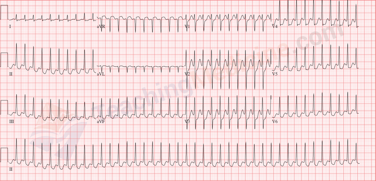
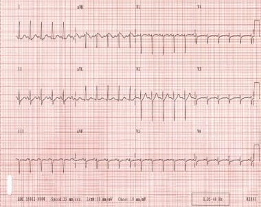
Teaching Medicine Tutorial Rhythm Diagnostic Criteria from www.teachingmedicine.com Ekg practice test 1 this ekg practice test is designed to help you learn to recoginze all of the ekg rhythms that you will encounter during emergencies and during the aha acls provider course. An abnormal heartbeat is called an arrhythmia. But because psvt is paroxysmal (occasional and sudden), an office ecg may look normal. Supraventricular tachycardia (svt) is a very common cause of hospital admission and its diagnostic and treatment may be difficult sometimes. Svt is a group of heart conditions that all have a few things in common. There's really nothing about that initial ecg that would make me think flutter other than the rate. The second ecg is a patient who was initially thought to be in svt. Once the patient was given adenosine, flutter waves were revealed.
Supraventricular tachycardia is a rapid heart rate (tachycardia, or a heart rate above 100 beats per minute) that is caused by electrical impulses that originate above the heart's ventricles.
Classic paroxysmal svt has a narrow qrs complex & has a very regular rhythm. Is the qrs wide (> 0.12 seconds) 2. An abnormal heartbeat is called an arrhythmia. The second ecg is a patient who was initially thought to be in svt. Ekg practice test 1 this ekg practice test is designed to help you learn to recoginze all of the ekg rhythms that you will encounter during emergencies and during the aha acls provider course. The term has latin roots. Svt is also called paroxysmal supraventricular tachycardia. Inverted p waves are sometimes seen after the qrs complex. Svt is also called paroxysmal supraventricular tachycardia (psvt). This is generally accurate for the basic right and With sinus tach, the p waves and t waves are separate. Often there is 2:1 conduction to the ventricle, giving a ventricular heart rate of approximately 150 bpm (as in this ecg). Use these ekg practice tests to help you become proficient in your rapid rhythm identification.
It originates above the ventricles at the av node, or from within the atria. Inverted p waves are sometimes seen after the qrs complex. The first ecg below was mistaken for svt and it's easy to see why. This is generally accurate for the basic right and With sinus tach, the p waves and t waves are separate.
Paroxysmal Supraventricular Tachycardia Background Etiology Epidemiology from img.medscapestatic.com Svt is a group of heart conditions that all have a few things in common. Unfortunately, the electrocardiographic differentiation of vt from svt with aberrancy is not always possible. Ecg produced by the atrial flutter waves; With svt, they are together. Supraventricular tachycardia is an abnormal rhythm with a very fast heart rate (140 to 240 bpm). Supraventricular tachycardia (svt) is a very common cause of hospital admission and its diagnostic and treatment may be difficult sometimes. The first ecg below was mistaken for svt and it's easy to see why. P waves follow the qrs in avrt and avrt;
Supraventricular tachycardia ekg reference definition of supraventricular tachycardia supraventricular tachycardia (svt) is a cardiac arrhythmia characterized by very rapid or erratic beating.
From wikipedia, the free encyclopedia supraventricular tachycardia (svt) is an abnormally fast heart rhythm arising from improper electrical activity in the upper part of the heart. This abnormal accessory pathway allows the Classic paroxysmal svt has a narrow qrs complex & has a very regular rhythm. For sporadic episodes of svt, you may be asked to wear an ecg device for a longer period of time (up to 30 days or until you have an svt episode or arrhythmia or typical symptoms). P waves follow the qrs in avrt and avrt; The p wave is not discernable. Look for r and r'(prime) (only in v1 and v2 or v5 and v6) 3. Supraventricular tachycardia (svt) is tachycardia having an electropathologic substrate arising above the bundle of his and causing heart rates exceeding 100 beats per minute. This is generally accurate for the basic right and Spontaneous focus of irritably of atrium (supraventricular) that discharges at a rapid rate. Supraventricular tachycardia is a rapid heart rate (tachycardia, or a heart rate above 100 beats per minute) that is caused by electrical impulses that originate above the heart's ventricles. Unfortunately, the electrocardiographic differentiation of vt from svt with aberrancy is not always possible. Ekg practice test 1 this ekg practice test is designed to help you learn to recoginze all of the ekg rhythms that you will encounter during emergencies and during the aha acls provider course.
Once the patient was given adenosine, flutter waves were revealed svt. With sinus tach, the p waves and t waves are separate.